 Dihedral (Shanghai) Science and Technology Co., Ltd
- Home
-
Products
-
- Semiconductor crystal
-
Single crystal substrate
-
Multifunctional single crystal substrate
- Barium titanate (BaTiO3)
- Strontium titanate (SrTiO3)
- Iron doped strontium titanate (Fe:SrTiO3)
- Neodymium doped strontium titanate (Nd:SrTiO3)
- Aluminium oxide (Al2O3)
- Potassium tantalum oxide (KTaO3)
- Lead magnesium niobate–lead titanate (PMN-PT)
- Magnesium oxide (MgO)
- Magnesium aluminate spinel (MgAl2O4)
- Lithium aluminate (LiAlO2)
- Lanthanu m aluminate (LaAlO3)
- Lanthanu m strontium aluminate (LaSrAlO4)
- (La,Sr)(Al,Ta)O3
- Neodymium gallate (NdGaO3)
- Terbium gallium garnet (TGG)
- Gadolinium gallium garnet (GGG)
- Sodium chloride (NaCl)
- Potassium bromide (KBr)
- Potassium chloride (KCl)
-
Multifunctional single crystal substrate
-
Functional crystal
- Optical window
- Scintillation crystal
-
Laser crystal
- Rare earth doped lithium yttrium fluoride (RE:LiYF4)
- Rare earth doped lithium lutetium fluoride (RE:LiLuF4)
- Ytterbium doped yttrium aluminium garnet (Yb:YAG)
- Neodymium doped yttrium aluminium garnet (Nd:YAG)
- Erbium doped yttrium aluminium garnet (Er:YAG)
- Holmium doped yttrium aluminium garnet (Ho:YAG)
- Nd,Yb,Er,Tm,Ho,Cr,Lu Infrared laser crystal
- N* crystal
- Metal single crystal
- Material testing analysis
- Material processing
- Scientific research equipment
-
-
Epitaxial Wafer/Films
-
Inorganic epitaxial wafer/film
- Gallium Oxide epitaxial wafer (Ga2O3)
- ε - Gallium Oxide (Ga2O3)
- Platinum/Titanium/Silicon Dioxide/Silicon epitacial wafer (Pt/Ti/SiO2/Si)
- Lithium niobate thin film epitaxial wafer
- Lithium tantalate thin film epitaxial wafer
- InGaAs epitaxial wafer
- Gallium Nitride(GaN) epitaxial wafer
- Epitaxial silicon wafer
- Yttrium Iron Garnet(YIG) epitaxial wafers
- Fullerenes&Fullerols
- ε-Gallium Oxide(Ga2O3)
- Indium Arsenide (InAs) epitaxial wafer
- InGaAs and other compound epitaxial wafers
- Periodic polarization of lithium niobate PPLN and lithium tantalate PPLT
-
Inorganic epitaxial wafer/film
- Functional Glass
- Fine Ceramics
-
2-D material
- 2-D crystal
-
Layered transition metal compound
- Iron chloride (FeCl2)
- Niobium sulfide (NbS3)
- Gallium telluride iodide (GaTeI)
- Indium selenide (InSe)
- Copper indium phosphide sulfide (CuInP2S6)
- Tungsten sulfide selenide (WSSe)
- Iron germanium telluride (Fe3GeTe2)
- Nickel iodide (NiI2)
- Iron phosphorus sulfide (FePS3)
- Manganese phosphorus selenide (MnPSe3)
- Manganese phosphorus sulfide (MnPS3)
- Interface thermal conductive materials
-
Epitaxial Wafer/Films
-
-
High-purity element
- Non-metallic
-
Metal
- Scandium (Sc)
- Titanium (Ti)
- Indium (In)
- Gallium (Ga)
- Bismuth (Bi)
- Tin (Sn)
- Zinc (Zn)
- Cadmium (Cd)
- Antimony (Sb)
- Copper (Cu)
- Nickel (Ni)
- Molybdenum (Mo)
- Aluminium (Al)
- Rhenium (Re)
- Hafnium (Hf)
- Vanadium (V)
- Chromium (Cr)
- Iron (Fe)
- Cobalt (Co)
- Zirconium (Zr)
- Niobium (Nb)
- Tungsten (W)
- Germanium (Ge)
- Iron(Fe)
-
Compound raw materials
-
Oxide
- Tungsten Trioxide (WO3)
- Hafnium Dioxide (HfO2)
- Ytterbium Oxide (Yb2O3)
- Erbium Oxide (Er2O3)
- Lanthanu m Oxide (La2O3)
- Cerium Dioxide (CeO2)
- Tin Dioxide (SnO2)
- Niobium Oxide (Nb2O3)
- Zirconium Dioxide (ZrO2)
- Zinc Oxide (ZnO)
- Copper Oxide (CuO)
- Magnetite (Fe3O4)
- Titanium Dioxide (TiO2)
- Samarium (III) oxide (Sm2O3)
- Silicon Dioxide (SiO2)
- Aluminum Oxide (Al2O3)
- Gallium Oxide Ga2O3(Powder)
- Sulfide
- Fluoride
- Nitride
- Carbide
-
Halide
- Gallium Chloride (GaCl3)
- Indium Chloride (InCl3)
- Aluminum Chloride (AlCl3)
- Bismuth Chloride (BiCl3)
- Cadmium Chloride (CdCl2)
- Chromium Chloride (CrCl2)
- Chromium Chloride Hydrate (CrCl2(H2O)n)
- Copper Chloride (CuCl)
- Copper Chloride II (CuCl2)
- Cesium Chloride (CsCl)
- Europium Chloride (EuCl3)
- Europium Chloride Hydrate (EuCl3.xH2O)
- Magnesium Chloride (MgCl2)
- Sodium Chloride (NaCl)
- Nickel Chloride (NiCl2)
- Indium Chloride (InCl3)
- Indium Nitrate Hydrate (In(NO3).xH2O)
- Rubidium Chloride (RbCl3)
- Antimony Chloride (SbCl3)
- Samarium Chloride (SmCl3)
- Samarium Chloride Hydrate (SmCl3.xH2O)
- Scandium Chloride (ScCl3)
- Tellurium Chloride (TeCl3)
- Tantalum Chloride (TaCl5)
- Tungsten Chloride (WCl6)
- Aluminum Bromide (AlBr3)
- Barium Bromide (BaBr2)
- Cobalt Bromide (CoBr2)
- Cadmium Bromide (CdBr2)
- Gallium Bromide (GaBr3)
- Gallium Bromide Hydrate (GaBr3.xH2O)
- Nickel Bromide (NiBr2)
- Potassium Bromide (KBr)
- Lead Bromide (PbBr2)
- Zirconium Bromide (ZrBr2)
- Bismuth Bromide (BiBr4)
- Bismuth Iodide (BiI3)
- Calcium Iodide (CaI2)
- Gadolinium Iodide (GdI2)
- Cobalt Iodide (CoI2)
- Cesium Iodide (CsI)
- Europium Iodide (EuI2)
- Lithium Iodide (LiI)
- Lithium Iodide Hydrate (LiI.xH2O)
- Gallium Iodide (GaI3)
- Gadolinium Iodide (GdI3)
- Indium Iodide (InI3)
- Potassium Iodide (KI)
- Lanthanu m Iodide (LaI3)
- Lutetium Iodide (LuI3)
- Magnesium Iodide (MgI2)
- Sodium Iodide (NaI)
-
Oxide
-
High-purity element
-
-
Sputtering Target
-
Metal target material
- Gold (Au(T))
- Silver (Ag(T))
- Platinum (Pt(T))
- Palladium (Pd(T))
- Ruthenium (Ru(T))
- Iridium (Ir(T))
- Aluminium (Al(T))
- Copper (Cu(T))
- Titanium (Ti(T))
- Nickel (Ni(T))
- Chromium (Cr(T))
- Cobalt (Co(T))
- Iron (Fe(T))
- Manganese (Mn(T))
- Zinc (Zn(T))
- Vanadium (V(T))
- Tungsten (W(T))
- Hafnium (Hf(T))
- Niobium (Nb(T))
- Molybdenum (Mo(T))
- Lanthanu m (La (T))
- Cerium (Ce (T))
- Praseodymium (Pr (T))
- Neodymium (Nd (T))
- Samarium (Sm (T))
- Europium (Eu (T))
- Gadolinium (Gd (T))
- Terbium (Tb (T))
- Dysprosium (Dy (T))
- Holmium (Ho (T))
- Erbium (Er (T))
- Thulium (Tm (T))
- Ytterbium (Yb (T))
- Lutetium (Lu (T))
- Alloy target material
- Semiconductor target material
-
Oxide target material
- Aluminum Oxide (Al2O3(T))
- Silicon Dioxide (SiO2(T))
- Titanium Dioxide (TiO2(T))
- Chromium Oxide (Cr2O3(T))
- Nickel Oxide (NiO(T))
- Copper Oxide (CuO(T))
- Zinc Oxide (ZnO(T))
- Zirconium Oxide (ZrO2(T))
- Indium Tin Oxide (ITO(T))
- Indium Zinc Oxide (IZO(T))
- Aluminum Doped Zinc Oxide (AZO(T))
- Cerium Oxide (CeO2(T))
- Tungsten Trioxide (WO3(T))
- Hafnium Oxide (HfO2(T))
- Indium Gallium Zinc Oxide (IGZO(T))
- Nitride target material
- Sulfide target material
-
Antimony tellurium selenium boron target material
- Magnesium Boride (MgB2(T))
- Lanthanu m Hexaboride (LaB6(T))
- Titanium Diboride (TiB2(T))
- Zinc Selenide (ZnSe(T))
- Zinc Antimonide (Zn4Sb3(T))
- Cadmium Selenide (CdSe(T))
- Indium Telluride (In2Te3(T))
- Tin Selenide (SnSe(T))
- Germanium Antimonide (GeSb(T))
- Antimony Selenide (Sb2Se3(T))
- Antimony Telluride (Sb2Te3(T))
- Bismuth Telluride (Bi2Te3(T))
-
Metal target material
-
Sputtering Target
-
- Services
- Media
- Partner
- Contact Us
- About
- Home
- Research Assistance
- Materials Analysis
Materials Analysis

| Material testing and analysis are very important in scientific research and engineering practice. Through testing and analysis, we can obtain accurate data and reliable results, providing a reliable basis for material selection, design, and application, and promoting the development of science and technology. |
| PtCat | Research Assistant cooperatively has a team of Shanghai Silicate Institute of the Chinese Academy of Sciences and Donghua University accumulated in the field of material research for more than 60 years, has more than 30 sets of various large-scale precision instruments and equipment, and provides (especially good at) professional and efficient testing and analysis of the composition, structure (crystal structure, molecular structure, and microstructure), thermal, mechanical, electrical and optical properties of inorganic nonmetals, I am very willing to work with researchers to solve the "difficult and complex problems" in material research. |
Main analyzing items:
| Index | Equipment | Description |
| 1 | High-power rotating target X-ray diffractometer | Grazing incidence XRD, residual stress, conventional XRD scanning, qualitative and quantitative phase analysis, crystallinity, accurate calculation of lattice parameters, grain size (microstrain), Rietveld structure refinement, etc. |
| 2 | High-power rotating target X-ray diffractometer | Grazing incidence XRD, conventional XRD scanning, qualitative and quantitative phase analysis, crystallinity, accurate calculation of lattice parameters, grain size (microstrain), Rietveld structure refinement, etc. |
| 3 | High-resolution powder X-ray diffractometer | Conventional XRD scanning, qualitative and quantitative phase analysis, crystallinity, accurate calculation of lattice parameters, grain size (microstrain), Rietveld structure refinement, etc. |
| 4 | Micro-focused two-dimensional X-ray diffractometer | Micro-area XRD analysis, residual stress determination, texture determination, high-throughput XRD characterization, etc. |
| 5 | In-situ X-ray diffractometer | XRD under different atmospheres and temperatures; in-situ chemical reaction and electrocatalytic XRD studies; study of crystal structure evolution under electric fields. |
| 6 | High-resolution X-ray diffractometer | 1. Grazing incidence diffraction (GID) for precise analysis of composition, order, and orientation in multilayer films; 2. X-ray reflectivity analysis (XRR) for precise analysis of thickness, density, and roughness in polycrystalline and single-crystal multilayer films; 3. High-resolution XRD (HRXRD) for rocking curve, reciprocal space mapping analysis in high-quality epitaxial films and single-crystal materials; 4. Annealing studies and high-resolution XRD characterization at high temperatures for epitaxial films and other materials. |
| 7 | Micro-focused rotating target single-crystal X-ray diffractometer | 1. Characterization of atomic coordinates, bond lengths, bond angles, configurations, thermal vibrations, and electron distributions in single crystals; 2. Characterization of microstructures such as atomic occupancy, defects, and bonding interactions; 3. Structural analysis of single crystals at medium and low temperatures. |
| 8 | Laser scanning confocal Raman spectroscopy | 1. Microscopic Raman spectroscopy measurements of powders, bulk materials, liquids, etc., and fluorescence spectroscopy measurements (473nm-1000nm); 2. Dispersive multi-point, line, and area scanning and confocal depth scanning for compositional distribution, stress distribution, etc.; 3. In-situ Raman spectroscopy measurements under temperature variations, in-situ electrocatalysis, and in-situ measurements in lithium (empty) batteries; 4. Polarized Raman spectroscopy measurements; 5. Raman spectroscopy characterization of large-sized samples; 6. Coupling with atomic force microscopy to simultaneously obtain morphological and compositional information for tip-enhanced Raman scattering (TERS). |
| 9 | Upright/inverted atomic force microscope | 1. Obtaining three-dimensional surface morphology of samples under ambient conditions without causing damage; 2. Various imaging modes including lateral force, phase, electrostatic force, piezoelectric force, and surface potential for characterization of dispersed multi-point, line, and area electrical/magnetic/mechanical properties (PFM/MFM/STM, etc.); 3. AFM characterization under high |
| 10 | Three-dimensional X-ray microscope | Two-dimensional tomography scanning and three-dimensional non-destructive imaging for studying microstructures and defects of samples, including morphology, porosity, cracks, etc. |
| 11 | High-resolution X-ray microscope | 1. Defect analysis: distribution of microcracks in materials, bonding degree of the matrix, the morphology of interfaces, local fiber orientation and thickness, particle size and shape, simulation of nucleation, growth, and coalescence processes. 2. Three-dimensional morphology analysis: characterization of pore size, distribution, types, and twisting of fibers, qualitative and quantitative characterization of microstructure parameters such as porosity, pore connectivity, and pore size, and 3D reconstruction of materials. |
| 12 | Field-emission scanning electron microscope (FE-SEM) | Surface or cross-section morphological observation of solid samples, fibers, films, etc.; observation and size analysis of micro/nano-particles, pores, etc.; analysis of element composition and distribution in micro-areas of the sample. |
| 13 | X-ray photoelectron spectroscopy (XPS) | XPS is primarily used for the qualitative and semi-quantitative analysis of the types, chemical states, chemical environments, and relative concentrations of elements within approximately 10 nm of a solid sample surface. It has wide applications in various disciplines related to solid materials, including polymers, metals, semiconductors, and thin films. |
| 14 | Field-emission transmission electron microscope (FE-TEM) | Analysis of microstructures within materials; analysis of element composition and distribution in micro-areas of the sample; analysis of material microstructures under heating or freezing conditions; in situ electrical and mechanical property testing of one-dimensional nanomaterials. |
| 15 | Field-emission transmission electron microscope (FE-TEM) | Analysis of microstructures within materials; analysis of element composition and distribution in micro-areas of the sample; TEM and STEM mode for three-dimensional reconstruction. |
| 16 | Transmission electron microscope (TEM) | Analysis of crystal defects, grain boundaries, phase boundaries, and other microstructures in metallic materials; analysis of morphological structures in polymer and composite materials; observation of particle shapes and size analysis; observation of shapes of chromosomes, ribosomes, proteins, hemoglobin, bacteria, etc. |
| 17 | Environmental scanning electron microscope (ESEM) | Surface morphology analysis of metallic, ceramic, polymer, and composite materials; surface morphology analysis of biological or aqueous samples (observation and analysis of samples without surface treatment); observation and size analysis of particles, porous materials, or fibers; qualitative and semi-quantitative analysis of surface micro-area composition of solid samples. |
| 18 | Scanning electron microscope (SEM) | Surface morphology observation of solid samples; analysis of fracture morphology and internal structure of materials; observation of particle or fiber shapes and size analysis. |
| 19 | Scanning probe microscope (SPM) | Analysis of surface morphology and phase composition of materials; analysis of various defects and contamination on material surfaces; study of surface mechanical properties; study of surface electrical and magnetic properties. |
| 20 | X-ray diffractometer (XRD) | Qualitative or quantitative phase analysis; crystal structure analysis; crystallinity determination (multiple peak separation method); grain orientation determination; study of phase transitions with temperature variation |
| 21 | Small-angle X-ray scattering instrument | Analysis of particle size, shape, and distribution of dispersed phases (particles, microcrystals, platelets, fillers, pores, and domains); analysis of particle dispersion state; study of interface structures; characterization of particle morphology in solution. |
| 22 | 600MHz Nuclear Magnetic Resonance (NMR) | Structural analysis of organic compounds; molecular structure analysis of natural products; analysis of biomolecular and protein structures; composition and chain structure analysis of polymer materials. |
| 23 | 400MHz Nuclear Magnetic Resonance (NMR) | Structural analysis of organic compounds and natural products; chain structure analysis of polymers; study of aggregation structures and polymer chain dynamics; study of interactions between polymer chains and compatibility of polymer blends. |
| 24 | Fourier Transform Infrared Spectrometer | Qualitative analysis of compounds; chain structure analysis of polymers; analysis of composition in mixtures; study of intermolecular interactions. |
| 25 | Fourier Transform Infrared Microscope | Qualitative analysis of trace samples; analysis of single fibers; analysis of composition distribution in micro-areas. |
| 26 | Gas Chromatography-Mass Spectrometry (GC-MS) | Structural analysis of organic compounds; qualitative and quantitative analysis of volatile organic mixtures; qualitative analysis of volatile organic compounds in water samples or solid samples; chain structure analysis of polymers; study of thermal degradation mechanisms of polymers. |
| 27 | Ultraviolet-Visible Spectrophotometer | Structural analysis of small molecule compounds; analysis of small molecule compound composition; transmission analysis of thin film samples; reflectance and transmission analysis of solutions, emulsions, or solid powders. |
| 28 | Inductively Coupled Plasma Emission Spectrometer (ICP-OES) | Determination of the content of more than 70 metallic and non-metallic elements in inorganic or organic samples, such as Cu, Pb, Cd, Cr, Ni, Zn, As, Fe, Bi, Ca, P, K, Mg, Al, Si, Sc, Sn, Mn, Zr, Ag, Ba, etc. |
| 29 | Elemental Analyzer | Determination of carbon, hydrogen, nitrogen, oxygen, and sulfur content in organic samples. |
| 30 | Inductively Coupled Plasma Mass Spectrometry (ICP-MS) | Determination of various metallic and non-metallic elements in inorganic or organic samples. |
| 31 | Glow Discharge Spectrometer (GDS) | Chemical element content analysis in matrices and coatings (films); elemental depth quantification for heat-treated workpieces (carburizing, nitriding); chemical element analysis in matrices covered by conductive or non-conductive coatings (films); analysis of chemical elements in solid samples covered by conductive or non-conductive coatings (films). |
| 32 | Steady-state/Transient Fluorescence Spectrometer | Chemical structure analysis of fluorescent substances; quantitative analysis of components with fluorescent properties; measurement of fluorescence quantum yield; determination of fluorescence lifetime and phosphorescence lifetime; time-resolved fluorescence spectroscopy; polarization properties of fluorescence. |
| 33 | Laser Micro-Raman Spectrometer | Determination of particle size and distribution of micelles, and colloidal particles; determination of the average molecular weight of polymers; study of molecular chain conformation and interactions with solvent molecules in polymer solutions; study of conformation and aggregation processes of proteins and polysaccharides. |
| 34 | Advanced Rotational Rheometer (ARES) | Determination of steady-state and dynamic rheological parameters for thermoplastic polymer melts; determination of rheological parameters for polymer solutions and other low-viscosity fluids; determination of rheological properties for shear-thinning fluids; determination of dielectric properties of polymers; determination of tensile properties of polymers. |
| 35 | Laser Micro-Raman Spectrometer (Micro-Raman) | Analysis of the chemical structure of materials (non-destructive qualitative analysis); analysis of aggregate structure, phase transformation, and defects; analysis of surface composition distribution and depth distribution; study of changes in the polymer structure, compatibility, stress relaxation, and interactions. |
| 36 | Gel Permeation Chromatography-Light Scattering Detector | Determination of absolute molecular weight and its distribution for polymers soluble in tetrahydrofuran. |
| 37 | Cryo-ultramicrotome | Preparation of semi-thin and ultra-thin sections, providing perfectly flat sections for optical microscopy, transmission electron microscopy, scanning electron microscopy, and atomic force microscopy. |
| 38 | Precision Ion Polishing System | Sample thinning and polishing for TEM samples of metals, ceramics, and semiconductors. |
| 39 | Microwave Digestion System | Sample pretreatment for ICP-OES, capable of digesting a wide range of organic or inorganic samples |
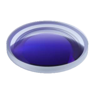
Special cold working of plane mirror and spherical mirror

1. Parameters of spherical mirror
General: radius of curvature (R), number of apertures (n), local aperture (△ n), finish (b), eccentricity (linear deviation C), angular deviation β)。
Special: specific dimensional tolerances.
2. Parameters of plane mirror
General, flatness refers to the number of apertures (n), local aperture (△ n), parallelism (/ /), angle (∠), perpendicularity (⊥), and fineness (B).
Special: specific dimensional tolerances.
| Ø 50 Ultra wide angle lens | Ø 70 glued objective | Ø 150 Beam splitter |
| 0.4mm Angle second level micro prism | 30_ Bevel cone lens | Step mirror |
| Φ 250 Spherical mirror | Ø 250 Spherical lens | Ø 300 Long lens |
| Wide angle mirror (multi sphere) | Conical lens | Hole positioning step mirror |
| Hexagonal quartz rod | 24x15x218 hexagonal quartz inclined rod | Angle rod mirror |
| Scanning glued prism group | Elliptical plane reflector | Octagonal strand prism (high speed scanning mirror) |
| Rod mirror of various sizes and precision | Strip mirror with various specifications and accuracy | Polorizing Prism |

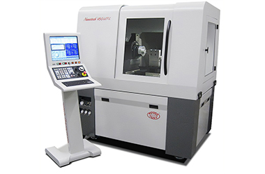
Moore Nanotech 450UPL Ultra-Precision Diamond Turning Lathe
The company has American Moore 450upl single point diamond superfinishing lathe and related testing equipment, which can provide external superfinishing services. The processing materials include pure aluminum, bronze, brass, germanium, nickel sulfide, zinc selenide, chalcogenide glass, plastics, etc. it can carry out ultra precision processing of reflective and transmissive optical elements with plane, cone, sphere, diffraction surface, paraboloid and other surface types. The surface roughness of the processing elements can reach nanometer level, and the maximum processing diameter is 400mm. 450upl ultra precision lathe has stronger processing capacity and is suitable for single point diamond processing and fixed-point micro grinding of optical components. 450upl can be used in optics, aerospace, national defense, consumer electronics, bearings, computer industry, etc. The slewing capacity is 450mm, which can be improved according to customer needs. B and C rotation axes can be selected to realize four axis linkage. When the spindle c-axis motion control is selected, the annular circular surface, biconical surface, off-axis aspheric surface and free-form surface can be processed by slow tool servo system (S3). Compared with grid processing, Moore nanotechnology's slow tool servo system can greatly reduce processing time and is easy to install and use. The surface finish and shape accuracy of machined parts are equivalent to those of rotationally symmetrical diamond machined parts.
It has American Moore 450upl single point diamond superfinishing lathe and related testing equipment, and can provide external superfinishing services. The processing materials include pure aluminum, bronze, brass, germanium, nickel sulfide, zinc selenide, chalcogenide glass, plastics, etc. it can carry out ultra precision processing of reflective and transmissive optical elements with plane, cone, sphere, diffraction surface, paraboloid and other surface types. The surface roughness of the processing elements can reach nanometer level, and the maximum processing diameter is 400mm.

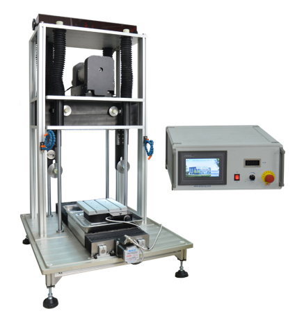
The company has a full-automatic diamond wire cutting machine, which is suitable for cutting materials with different hardness, such as ceramics, crystals, glass, metals, rocks, thermoelectric materials, infrared optical materials, composites and biomedical materials. It is mainly used to process large-scale precious materials, and the cutting size can reach 12 ". The full-automatic diamond wire cutting machine has high cutting accuracy and can meet the cutting of most materials. The full-automatic diamond wire cutting machine is a continuous cutting diamond wire cutting machine. After setting the cutting procedure, the sample is fed continuously without manual adjustment. The size accuracy of the cut sample is high, within ± 10% μ M.
Detailed parameters are as follows:
1. The main motor drives the diamond cutting line to move downward at a constant speed, and the material is fixed on the workbench to ensure the stability of cutting.
2. The workbench can be operated manually or through program control ˚ Rotation adjustment.
3. Pneumatic tensioning system and imported pneumatic components are adopted to make the tensioning force more stable and reliable.
4. PLC program control system and large touch screen make the operation simple and fast.
5. We can design tools and fixtures according to your needs.
Automatic Diamond Wire Cutting
1. Ceramic materials: alumina ceramics, zinc oxide ceramics, zirconia ceramics, target ceramics, honeycomb ceramics, semiconductor ceramics, conductive ceramics, non-conductive ceramics, etc;
2. Crystal materials: graphite, silicon crystal (solar polysilicon, monocrystalline silicon), sapphire, alumina crystal, infrared glass crystal, alumina crystal, silicon carbide crystal, cesium iodide crystal, etc;
3. Glass materials: chalcogenide glass, optical glass, quartz glass, infrared glass, glass tube, etc;
4. Metal materials: iron, aluminum, copper, titanium alloy, magnesium alloy and other metals and alloys, non-ferrous metals (zinc sulfide, ferrite), etc;
5. Composite materials: PVC board, carbon fiber composite, glass fiber composite, etc
6. Rock materials: precision cutting of natural rock, jade, meteorite, Peicui, agate and other high-value materials; Geological light slice, geological thin slice (sedimentary rock, magmatic rock, metamorphic rock, ore), etc.
7. Thermoelectric materials: bismuth telluride, lead telluride, silicon germanium alloy, etc
8. Infrared optical materials: zinc selenide, zinc sulfide, silicon, germanium and other crystals
9. Biomedical materials: bioplastic specimen sections (joint sections of human animal organs, jaw soft and hard tissues, implant observation, dental crown and bridge, teeth and other histological specimens); Joint sections of soft and hard tissues in orthopedics (fresh and hard tissues such as femur, hip joint and vertebral body, bone histological samples with implants, etc.); Cardiovascular and cerebrovascular stent sections, stone sections and other medical tissue sections.

